Choroidal neurofibromas in Neurofibromatosis 1
Choroidal neurofibromas in Neurofibromatosis 1
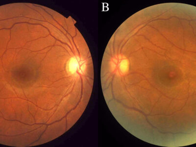
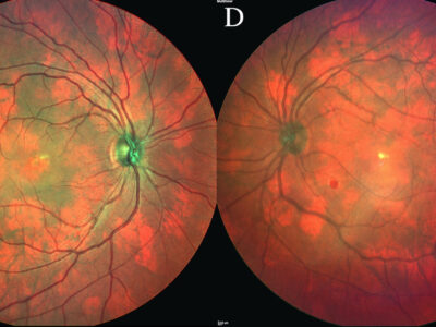
A 28-year-old female, with neurofibromatosis 1 (NF1), presented to the clinic with a history of reduced vision in the left eye. On examination, in addition to Lisch nodules in both eyes, a cataract was observed in the left eye. Dilated fundus ophthalmoscopy was unremarkable (Figure 1). With multicolour retinal imaging however, both eyes exhibited red patches distributed throughout the fundus (Figure 2). No retinal abnormalities were detected on optical coherence tomography or fluorescein angiography in either eye. Based on these findings, a diagnosis of choroidal neurofibromas was made. Choroidal neurofibromas are benign hamartomatous proliferation of neural crest derived cells of the choroid. The sensitivity of choroidal neurofibromas in NF1 is 97.5% compared to café au lait spots (98%) and Lisch nodules (86%). Of note, only 2% of NF1 patients present with Lisch nodules in the absence of choroidal neurofibromas whereas 13.75% of patients present with choroidal neurofibromas in the absence of Lisch nodules. As a result, screening for choroidal neurofibromas has been suggested as a highly sensitive non-invasive screening tool for detection of NF1. This case highlights that standard fundus examination is likely to miss an essential diagnostic feature of NF1 in the eye. Multicolour fundus imaging should be used in of NF1.


