OCT assessment of papilloedema
OCT assessment of papilloedema
Patient 1:
Background/indication: young lady with papilloedema secondary to IIH who needed urgent VP shunt to save her vision.
Description:
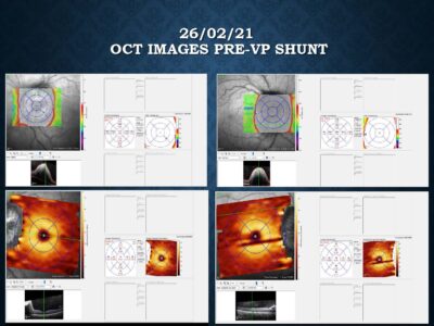
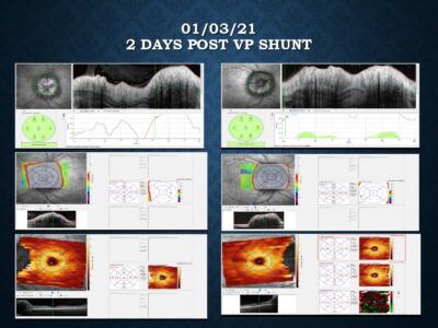
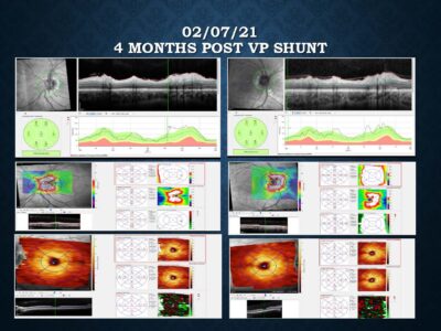
OCT optic disc volume scans pre and post VP shunt insertion.
1. 26/02/2021: vision threatening level of papilloedema (Frisen V) at initial presentation.
2. 04/03/2021 : significant regression in the level of papilloedema 3 weeks following VP shunt.
3. 02/07/2021 : resolution of papilloedema 4 months after VP shunt insertion.
Patient 2:
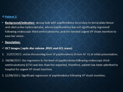
Background/indication: young lady with papilloedema secondary to tectal plate lesion and obstructive hydrocephalus, whose papilloedema has not significantly regressed following endoscopic third ventriculostomy ,and she needed urgent VP shunt insertion to save her vision.
Description:
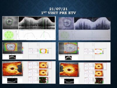
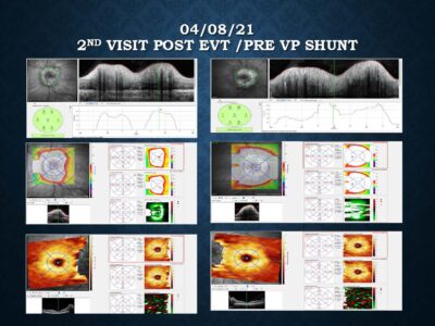
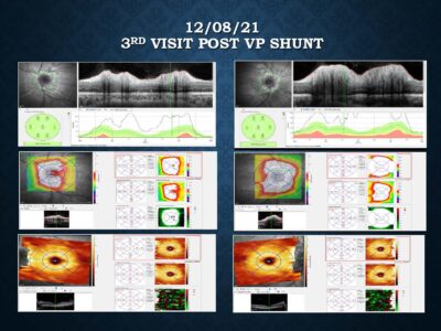
OCT images ( optic disc volume ,RNFL and GCL scans)
1. 21/07/2021: vision threatening level of papilloedema (Frisen IV- V) at initial presentation.
2. 04/08/2021: the regression in the level of papilloedema following endoscopic third ventriculostomy (ETV) was less than the expected, therefore, patient has been admitted to hospital for urgent VP shunt insertion.
3. 12/08/2021: Significant regression of papilloedema following VP shunt insertion.
Conclusion:
1. OCT has a major role in the diagnosis and monitoring of papilloedema and other different neuro ophthalmic conditions.
2. Optic disc OCT supports the clinical assessment of papilloedema severity alongside the clinical examination and formal visual field assessment.


